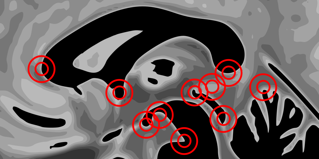
An open framework for evaluating correspondence in brain images and teaching neuroanatomy using anatomical fiducial placement
Preprint: https://www.biorxiv.org/content/10.1101/460675v2
Manuscript: http://dx.doi.org/10.1002/hbm.24693
Documentation: https://afids.readthedocs.io/en/latest/
- The AFID protocol is an anatomical fiducial placement protocol that has been validated and used for teaching at a number of local and BHG-related events including https://github.com/BrainhackWestern/BrainhackWestern.github.io/wiki/Tutorials.
- AFID placement is reproducible, not overtly manually intensive (20-40 minutes once trained), and more sensitive to local registration errors than standard voxel overlap measures.
- This protocol and study framework leverages open resources and tools, and has been developed with full transparency in mind so that others may freely use, adopt, and modify.
- 60+ raters trained to date.
- Lau JC, Parrent AG, Demarco J, Gupta G, Park PJ, Ferko K, Khan AR, Peters TM. A framework for evaluating correspondence between brain images using anatomical fiducials. bioRxiv. 2018. [ref]
- Lau JC, Parrent AG, Demarco J, Gupta G, Park PJ, Ferko K, Khan AR, Peters TM. AFIDs: an open framework for evaluating correspondence between magnetic resonance images of the human brain using fiducial placement. F1000 Research. Demo presented at INCF NeuroInformatics in Montreal, QC, Canada. 2018. [ref]
- Agile12v2016: Lau JC, MacDougall KW, Arango MF, Peters TM, Parrent AG, Khan AR: Ultra-High Field Template-Assisted Target Selection for Deep Brain Stimulation Surgery. World Neurosurg 103:531–537, 2017. [download] [ref]
- Colin27: Holmes CJ, Hoge R, Collins L, Woods R, Toga AW, Evans AC: Enhancement of MR Images Using Registration for Signal Averaging. J Comput Assist Tomogr 22:324–333, 1998. [download] [ref]
- MNI152NLin2009bAsym: Fonov V, Evans AC, Botteron K, Almli RR, McKinstry RC, Collins LL, et al: Unbiased average age-appropriate atlases for pediatric studies. Neuroimage 54:313–327, 2011. [download] [ref]
- OASIS1: Marcus DS, Fotenos AF, Csernansky JG, Morris JC, Buckner RL: Open Access Series of Imaging Studies: Longitudinal MRI Data in Nondemented and Demented Older Adults. J Cogn Neurosci 22:2677–2684, 2010. [download] [ref]
- 3D Slicer: Fedorov A, Beichel R, Kalpathy-Cramer J, Finet J, Fillion-Robin JC, Pujol S, et al: 3D Slicer as an image computing platform for the Quantitative Imaging Network. Magn Reson Imaging 30:1323–1341, 2012. [download] [ref]
- Talairach1957: Talairach J, David M, Tournoux P, Corredor H, Kvasina T: Atlas d’anatomie Stéréotaxique. Repérage Radiologique Indirect Des Noyaux Gris Centraux Des Régions Mésencephalosousoptique et Hypothalamique de l’homme. Paris, France: Masson & Cie, 1957
- Talairach1988: Talairach J, Tournoux P: Co-Planar Stereotaxic Atlas of the Human Brain: 3-D Proportional System: An Approach to Cerebral Imaging. ed 1, Thieme, 1988.
- Website for the AFID protocol as taught as a training workshop for BHG18
- Tutorial given as part of BHG18 [video]
- Download Slicer
- AFID validator (alpha stage); started at BHG18-Western
- Scicrunch (RRID:SCR_016623)


